
X-Ray Imaging in Animals: The Complete Guide to Getting Started

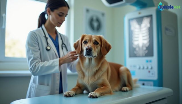
X-ray imaging is a painless diagnostic procedure that allows veterinarians to visualize bones, organs, and certain foreign objects inside an animal's body. This practical guide explains how the examination is performed, why it's prescribed, and how to prepare your animal properly so you can approach the x-ray with confidence and without stress.
1. What is X-Ray Imaging and What Is It Used For?
X-ray imaging uses radiation to produce images of an animal's internal structures. Dense tissues, such as bones, appear light on the image, while soft tissues and air appear darker. This examination helps the veterinarian confirm or rule out many hypotheses before reaching an accurate diagnosis.
It is used in both companion animals and livestock for a wide variety of situations: lameness, trauma, persistent cough, suspected ingestion of a foreign object, pregnancy monitoring, or routine health assessment before surgical intervention.
What X-Ray Imaging Can Reveal
- Fractures, dislocations, and bone abnormalities
- Certain tumors or abdominal masses
- Ingested foreign objects (dense materials)
- The size and shape of the heart and lungs
- The presence of urinary calculi in certain animals
- The approximate number of offspring in late pregnancy
2. When Does Your Veterinarian Recommend X-Ray Imaging?
The decision to perform x-ray imaging always depends on a prior clinical examination. Your veterinarian bases this decision on the symptoms observed and physical palpation findings.
| Symptom Observed | Value of X-Ray Imaging | Typical Urgency Level |
|---|---|---|
| Sudden lameness after trauma | Rule out fracture or dislocation | High |
| Chronic cough or shortness of breath | Examine lungs and heart | Moderate |
| Repeated vomiting | Detect a gastrointestinal foreign object | High |
| Difficulty urinating | Check for urinary calculi | High |
| Late pregnancy | Estimate number of offspring | Low to Moderate |
3. How Is the X-Ray Examination Performed?
X-ray imaging is performed at a veterinary clinic on a dedicated table. The animal is positioned carefully, sometimes with the help of straps or positioning aids, to obtain a clear image without causing injury.
- The veterinarian or veterinary assistant positions the animal in the appropriate stance for the area being examined.
- Gentle restraint is applied to prevent movement during image capture.
- The equipment emits a brief, painless burst of radiation to capture the image.
- Multiple images from different angles are often necessary for complete interpretation.
- The veterinarian then analyzes the images and explains the results to the owner.
In some cases, light sedation is offered if the animal is too stressed, in pain, or restless to remain still safely.
4. Preparing Your Animal for the Examination
Good preparation makes the veterinarian's work easier and reduces stress for your animal. A few simple steps ensure you arrive prepared on the day of the x-ray.
- Follow fasting instructions if sedation is planned
- Bring your animal calm, without excessive excitement before travel
- Use a secure carrier for small animals during transport
- Inform your veterinarian of any current medications
- Bring your animal's health records and results from previous examinations
5. Common Mistakes to Avoid
Certain habits delay diagnosis or unnecessarily complicate the examination. Here are the most common pitfalls encountered among first-time pet owners.
- Waiting too long before consulting when lameness or visible pain is present
- Feeding your animal before an examination when fasting was requested
- Neglecting proper handling at home, making the animal more difficult to manage at the clinic
- Interpreting images alone without your veterinarian's professional opinion
- Refusing sedation out of fear, when it actually ensures the examination is safe for a painful animal
6. After the X-Ray: Reading Results and Follow-Up Care
Once the images are obtained, your veterinarian takes time to explain what they observe in understandable terms. They may recommend additional tests if images alone are insufficient to reach a conclusion, such as ultrasound or blood work.
Depending on the results, treatment, immobilization, surgery, or simple monitoring may be recommended. It is essential to follow post-examination instructions, especially if sedation was used: your animal should remain calm while fully recovering from the procedure.
For any questions or to schedule this examination, it is recommended to consult the veterinarian directory to find a qualified professional with the necessary equipment near you.
Summary
- X-ray imaging is a painless procedure that visualizes bones, organs, and foreign objects
- It is prescribed following a clinical examination, never routinely or without cause
- Good preparation (fasting, calmness, information) facilitates the examination
- Light sedation is sometimes necessary for your animal's safety
- Results must always be explained and interpreted by a veterinarian
Frequently Asked Questions
Is x-ray imaging painful for my animal?
No, the examination itself is completely painless. The only possible discomfort comes from the restraint necessary to keep your animal still for a few seconds.
Do I need to fast my animal before x-ray imaging?
This depends solely on whether sedation or anesthesia is needed. Your veterinarian will provide specific fasting instructions based on the type of examination planned.
How long does an x-ray appointment take?
The actual image capture is very quick, but preparation, positioning, and sometimes sedation can extend the total visit duration. Your veterinarian will give you an estimate before the examination.
If you have any doubt about your animal's health, x-ray imaging remains a valuable veterinary tool for reaching a reliable and quick diagnosis.
Leave a comment
Be the first to comment on this article.

TunisieVet in your pocket 📱🐾 Access your veterinary services wherever you are. [ Download the app ]
Learn more