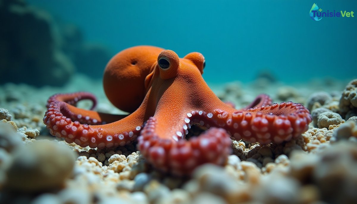
The common octopus changes color in less than a second because its chromatophores are activated directly by muscles connected to the central nervous system, without going through the slower hormonal pathway. This direct neuromuscular control allows an almost instantaneous reaction to a threat or to blend into the background.
A muscular system, not glandular
Each chromatophore is a sac of pigment (yellow, orange, red or brown) surrounded by around fifteen tiny radial muscles. When the brain sends a nerve impulse, these muscles contract and stretch the sac, exposing the pigment over an area up to 500 times larger; at rest, the muscles relax and the sac closes. As each cell is individually wired to the brain, Octopus vulgaris can modulate tens of thousands of chromatophores almost simultaneously, which would never be possible with a hormonal mechanism, which is too slow and diffuse.
Under this layer of pigments are two other types of cells which complete the palette: iridophores, stacks of reflective plates which produce metallic, bluish or greenish reflections by diffraction of light, and leucophores, cells diffusing white light to imitate the light background or reinforce the contrast of the patterns. The combination of the three layers allows the octopus to reproduce textures, zebra patterns or spots in a few tenths of a second.
A colorblind person who “sees” with the skin?
The paradox is documented by several studies: the eyes of Octopus vulgaris only have one type of photoreceptor, which should make it colorblind and unable to distinguish the colors of its environment. Yet he adjusts his hue with a precision that suggests chromatic perception. The most studied hypothesis is that opsins, photosensitive proteins usually confined to the retina, would also be present in the skin itself, allowing a form of cutaneous light detection that would help calibrate camouflage independently of ocular vision. This mechanism remains actively studied and is not fully elucidated.