Veterinary case
A dog's huge swollen belly: double uterine torsion
⚠️ This information is provided for awareness only and does not replace an examination by a veterinarian.
🤖 Summary produced automatically from the scientific publication cited at the bottom of the page.
An old intact female dog arrived with a hugely swollen belly: both horns of her uterus had twisted around each other.
What happened?
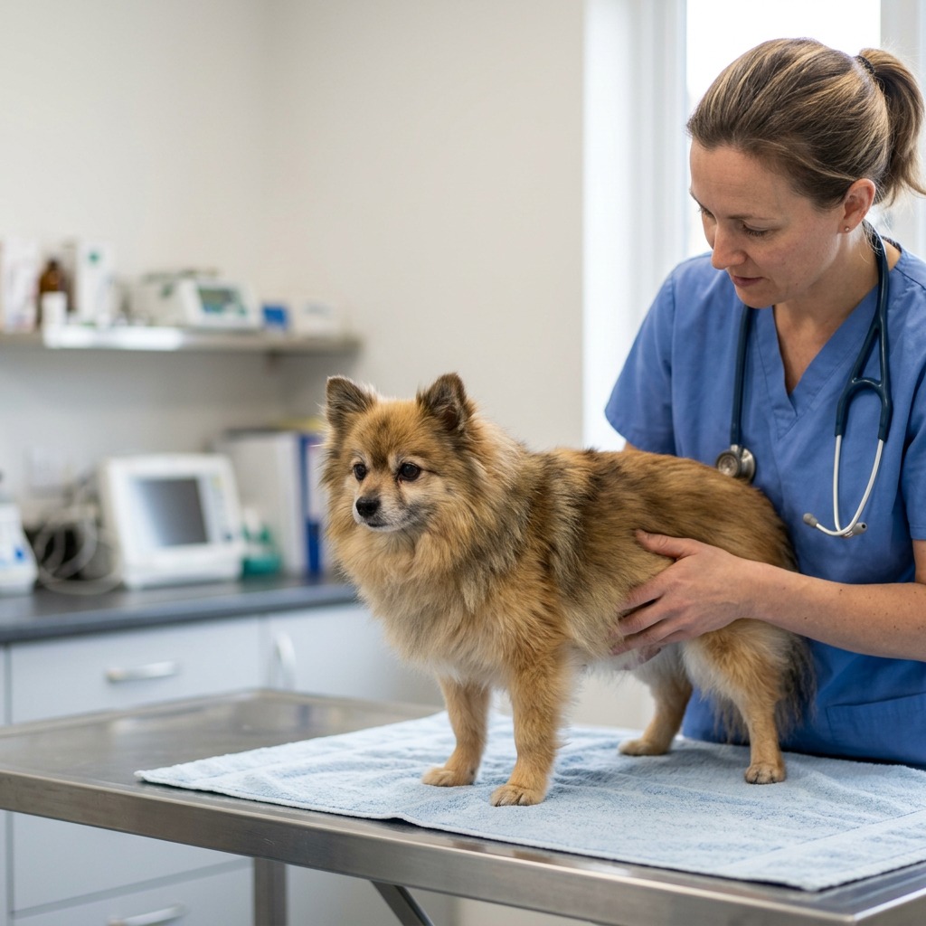
An 11-year-old intact female Pomeranian, who had never had puppies, was brought to the vet after 2 to 3 days of abdominal swelling, breathing difficulty, loss of appetite, vomiting, and low urine output. On examination, her belly was very distended, with a large mass felt inside and another mass visible at the vagina. The dog was lethargic and breathing fast.
What did the vet find?
Blood tests showed the kidneys were starting to fail, with abnormal blood salt levels and signs of severe inflammation in the body. X-rays and ultrasound showed two large masses in the belly, one on each side, with a little fluid around them, but without being able to tell exactly where they came from. A contrast CT scan showed that these masses actually came from both uterine horns, that blood flow to them was almost cut off, and that both horns were twisted on themselves, the right one far more twisted than the left. This allowed the diagnosis of bilateral uterine horn torsion to be made before surgery.
What did the vet do?
ℹ️ Treatment given to THIS animal, decided by its veterinarian. It is not a prescription: never apply it without veterinary advice.
The vet decided to operate directly without trying to untwist the uterus first, because this could have released toxic substances into the dog's blood. During surgery, he found that the right horn had rotated about three full turns around the left one, and the left horn had itself twisted about a quarter turn. The whole uterus, along with the ovaries and the vaginal mass, was removed in one piece.
What happened next?
The microscope examination showed the masses were benign uterine tumors, not cancer, but the right horn had suffered a lot from lack of blood supply with dead tissue areas. The dog recovered well from anesthesia and surgery and went home without complications. Nine months later, she was still doing well with no sign of recurrence.
Advice for the farmer
In an older intact female dog, rapid abdominal swelling with breathing difficulty and loss of appetite should raise concern: spaying female dogs that are not used for breeding helps avoid this kind of serious uterine complication.
Source
- Source
- PubMed Central
- Title
- Case Report: Computed tomographic diagnosis of asymmetric bilateral uterine horn torsion secondary to benign uterine leiomyomas in a non-gravid dog
- Authors
- Jeong E, Kim J, Kim Y, Eom K, Kim J.
- Licence
- CC BY