Veterinary case
A cat can't walk: a mass on the spine
⚠️ This information is provided for awareness only and does not replace an examination by a veterinarian.
🤖 Summary produced automatically from the scientific publication cited at the bottom of the page.
A young cat gradually loses the use of her hind legs, and scans reveal a mass on one vertebra.
What happened?
A 2-year-old spayed Scottish Fold cat had been less active for 2 weeks. Her hind legs grew weaker until she could no longer walk. She also struggled to urinate and defecate.
What did the vet find?
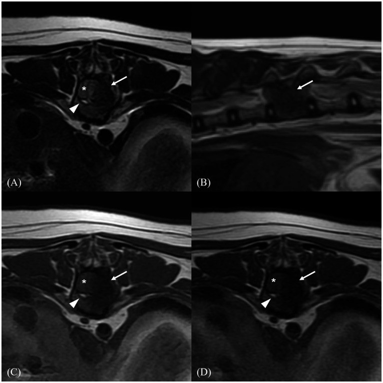
Blood and urine tests were normal. The cat had back pain and weak reactions in her hind legs. X-ray, CT and MRI showed a bony mass on a mid-back vertebra strongly squeezing the spinal cord. Tissue analysis showed a blood vessel malformation, with two labs giving related names.
What did the vet do?
ℹ️ Treatment given to THIS animal, decided by its veterinarian. It is not a prescription: never apply it without veterinary advice.
The surgeon opened the back and removed the abnormal bone pressing on the spinal cord. Samples were sent for analysis, and the area was covered with the cat's own fat.
What happened next?
After 7 days the cat could walk again, though weakly. By day 20 her gait was normal and she had regained control of urine and stool. No relapse in 75 days.
Advice for the farmer
Progressive hind-leg weakness in a young animal deserves a prompt exam: treated in time, the spinal cord can recover.
Source
- Source
- PubMed Central
- Title
- Case Report: Spinal arteriovenous fistula and vertebral angiomatosis in a cat-the role of imaging within the vascular malformation spectrum.
- Authors
- Oh S, Ahn S, Lee K, Yoon H.
- Year
- 2026
- DOI
- 10.3389/fvets.2026.1862231
- Licence
- CC BY