Veterinary case
Bovine membrane heals a cat's large open skin wound
⚠️ This information is provided for awareness only and does not replace an examination by a veterinarian.
🤖 Summary produced automatically from the scientific publication cited at the bottom of the page.
A young cat lost a large patch of back skin after treatment, then recovered thanks to an unusual technique borrowed from cattle.
What happened?
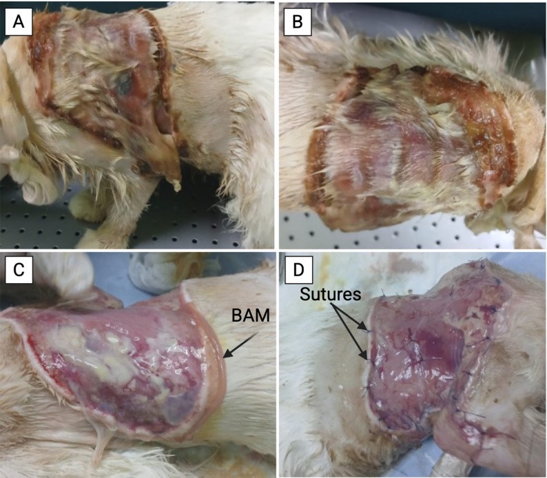
An eight-month-old cat received a subcutaneous fluid injection to treat a viral infection. A few days later, the skin on her back became wet, fragile and started sloughing off, with pus discharge. She was lethargic, feverish, and eating very little.
What did the vet find?
The vet found a large area of dead skin on the back and flank, covering about 12 to 15 percent of the body surface. The skin was wet, fragile and oozing pus, a sign of infection together with tissue death.
What did the vet do?
ℹ️ Treatment given to THIS animal, decided by its veterinarian. It is not a prescription: never apply it without veterinary advice.
The vet first gave antibiotics and cleaned the wound daily for a week to control infection. Once the wound was clean, he placed a membrane taken from the placenta of a healthy cow, preserved with propolis, over the raw area and stitched it in place. A month later, a second membrane was applied to complete healing, along with daily wound care and a cream to keep the wound moist.
What happened next?
The wound closed progressively, healing faster after each membrane application. Within a little over a month the new skin had fully covered the area, and after about two and a half months the fur had regrown normally with no infection or visible scarring. The cat regained her appetite and overall health.
Advice for the farmer
After any subcutaneous injection, watch the area closely in the following days: any redness, swelling or skin sloughing should be shown to the vet quickly.
Source
- Source
- PubMed Central
- Title
- Successful management of a large feline skin defect using xenogeneic bovine amniotic membrane: a case report.
- Authors
- Waleed A, Abu-Ahmed H, Kassem M, Elkhenany H.
- Year
- 2026
- DOI
- 10.1186/s12917-026-05389-9
- Licence
- CC BY