
Intestinal Parasites in Ruminants: Recognition and Management

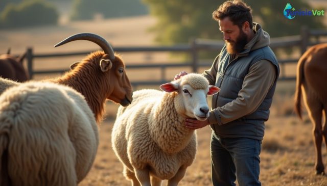
Intestinal parasites in ruminants rank among the leading causes of productivity losses in cattle, sheep, and goat farming. Diarrhea, weight loss, declining milk production: these signs often mislead farmers. This article explains how to identify parasitic infestation, match anthelmintic treatment to parasite lifecycles, and build a preventive schedule suited to Mediterranean climate conditions without accelerating antiparasitic resistance.
1. Understanding the main intestinal parasites of ruminants
Worms in goats, sheep, and cattle belong primarily to three major families. Gastrointestinal strongyles (including the notorious Haemonchus contortus, a blood-feeder) colonize the abomasum and small intestine. Liver flukes, both large and small, lodge in the bile ducts after passing through an intermediate aquatic host—the snail. Coccidia, conversely, are not worms but protozoans affecting mainly young animals.
A lifecycle dependent on climate
The development of infective larvae on pasture depends heavily on humidity and temperature. In Mediterranean climate regions, the rainy periods of late autumn and spring favor larval hatching and survival on grass, whereas dry summer heat slows their development but allows resistant forms to persist in soil.
2. Signs of parasitic infestation to monitor in the herd
Signs of parasitic infestation in livestock are often subtle at first, delaying diagnosis. Regular observation of the herd helps identify animals at risk before conditions worsen.
- Persistent diarrhea, sometimes tinged with blood in cases of heavy strongyle or coccidia burden.
- Progressive weight loss despite maintained appetite or normal forage intake.
- Anemia, visible as pale mucous membranes in the eye or gums—characteristic of haemonchosis infections.
- Submandibular edema ("bottle jaw"), a classic sign of severe chronic parasitosis in sheep and goats.
- Production decline: slower weight gain in young stock, drop in milk yield in adult females.
- Rough, dull coat and overall loss of vigor in the herd.
An animal isolated from the group, lying down more frequently than usual, or showing an distended abdomen warrants close examination—even fecal sampling for coproscopic analysis.
3. Diagnose before treating: fecal examination
Treating a herd without knowing which parasite is responsible or the severity of infestation amounts to wasting product and accelerating antiparasitic resistance. Coproscopic analysis, performed by a veterinary laboratory on fecal samples, counts eggs per gram of feces and guides treatment selection.
This examination proves particularly valuable:
- before spring turnout, to assess residual parasitic burden;
- when unexplained clinical signs appear in multiple animals;
- after treatment, to verify real-world efficacy.
4. Match anthelmintic treatment to parasite lifecycle
Effective anthelmintic treatment in ruminants hinges on the right drug, at the right time, for the right animal. The most commonly used drug families are benzimidazoles, macrocyclic lactones, and imidazothiazoles—each with its own spectrum of action and limitations.
| Period | Dominant parasitic risk | Recommended action |
|---|---|---|
| Late autumn | Strongyles, liver fluke after rains | Coproscopic check; targeted treatment if needed |
| Winter | Reduced but non-zero risk in wet zones | Monitor; avoid waterlogged pastures |
| Spring | Larval activity resumes; coccidiosis in young stock | Targeted treatment before intensive grazing |
| Dry summer | Low larval development on pasture | Limit unnecessary blanket treatments |
Treatment should never be routine for the entire herd. Targeting animals most severely affected, identified by clinical examination or coproscopy, preserves drug efficacy long-term.
5. Build a realistic deworming schedule for Mediterranean climate
A deworming calendar for ruminants in Tunisia must account for two risk peaks tied to autumn and spring rains, separated by a dry summer less conducive to larval development.
- Before spring turnout: systematic coproscopic check; targeted treatment of positive animals.
- Late summer, before autumn rains: assess body condition of the herd and weaned young stock, often more vulnerable.
- At housing or during winter confinement: verify absence of reinfestation before animals are grouped.
- Throughout the year: close monitoring of young animals, pregnant females in late gestation, and weakened animals—all more susceptible to parasites.
Wet grazing areas near water points or irrigated zones demand heightened vigilance due to liver fluke risk linked to the presence of snail intermediate hosts.
6. Prevent without over-treating: non-drug management tools
Internal parasite prevention in livestock rests not solely on anthelmintic products. Several management practices durably reduce parasitic pressure.
- Pasture rotation: alternating paddocks and, where possible, animal species (cattle and small ruminants harbor different strongyles) breaks the parasite cycle.
- Cutting or mixed grazing: allowing a paddock several weeks of rest reduces the burden of infective larvae.
- Avoid close grazing: larvae concentrate in the first centimeters of grass; not allowing animals to graze too short limits ingestion.
- Watering management: avoid marshy or poorly drained zones—breeding grounds for liver fluke.
- Balanced nutrition: well-fed animals resist moderate parasitic burden better.
7. Antiparasitic resistance: mistakes to avoid at all costs
Antiparasitic resistance in livestock advances when the same drugs are used repeatedly and without reasoning. Several classic errors accelerate this phenomenon.
- Routinely treating the entire herd without distinguishing between affected and healthy animals.
- Under-dosing by eyeballing weight rather than weighing or measuring with a tape.
- Always using the same drug family from year to year without planned rotation.
- Treating then immediately moving animals to clean pasture, which favors selection of resistant survivors.
- Ignoring results of ineffective treatment instead of having a professional investigate the cause.
Leaving part of the herd untreated (the least affected animals) maintains a population of parasites sensitive to available drugs, limiting resistance spread.
When facing persistent signs or uncertainty about protocol choice, professional guidance remains essential. A veterinarian specializing in livestock can establish precise diagnosis and tailor anthelmintic treatment to actual herd circumstances, whether managing cattle, sheep, or goats.
Summary
- Observe the herd regularly: diarrhea, weight loss, and anemia are early warning signs.
- Confirm diagnosis with fecal examination before any targeted anthelmintic treatment.
- Align deworming schedule with rainy periods—the main drivers of parasite cycles in Mediterranean climates.
- Combine pasture rotation and watering management with drug treatment.
- Avoid blanket herd treatment to preserve drug efficacy over the long term.
Frequently asked questions
How often should you deworm a ruminant herd?
No single frequency applies everywhere: it depends on climate, grazing system, and coproscopic analysis results. Targeted treatment based on observation and diagnosis beats a fixed calendar applied blindly.
How can you tell if your animals have worms?
Suggestive signs include diarrhea, weight loss, pale mucous membranes, and declining milk production. Only laboratory fecal analysis confirms the type and intensity of infestation.
Can you prevent intestinal parasites without medication?
Pasture rotation, avoiding short grazing, and sound watering management significantly reduce risk. These measures don't replace treatment when infestation is confirmed but limit blanket reliance on anthelminthics.
Managing intestinal parasites in ruminants demands method over routine. By combining careful observation, laboratory diagnosis, and sound grazing practices, farmers protect their herd's health sustainably while preserving the future effectiveness of treatments.
Leave a comment
Be the first to comment on this article.